
- The basic layout of the light microscope as consisting of a microscope objective and an eye-piece, and their properties such as aberrations, magnification, numerical aperture, and field of view.
- Different methods for obaining contrast in a microscope, such as absorption, fluorescence, and phase-contrast methods.
- Different image-quality measures such as resolution, contrast, signal-to-noise ratio (SNR), modulation transfer function (MTF), sampling density, field of view, and depth of field.
- Layout of the illumination systems, particularly Köhler illumination for transmission and epi-fluorescence microscopy, and how the illumination system can be used to optimize image quality.
- How properties of the microscope and illumination system affect image quality such as resolution and contrast, mainly via Fourier methods including point-spread functions (PSF) and optical transfer functions (OTF, MTF).
- How the choice of detector affects image quality measures such as signal-to-noise (SNR) and sampling. How to sample to avoid loss of information and artefacts. Some microscope photometry.
- The basic layout for confocal microscopy and hence three-dimensional imaging, including resolution and sampling in different dimensions.
- The basics of nanoscopy and imaging beyond the classical resolution limit.
SK2500 Physics of Biomedical Microscopy 6.0 credits
This course will be discontinued.
Last planned examination: Autumn 2026
Decision to discontinue this course:
No information inserted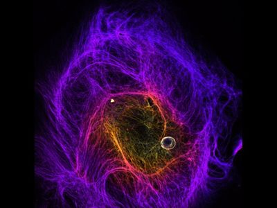
The course covers the physics and technology of microscopic imaging methods, with special emphasis on applications within the life sciences.
Information per course offering
Course offerings are missing for current or upcoming semesters.
Course syllabus as PDF
Please note: all information from the Course syllabus is available on this page in an accessible format.
Course syllabus SK2500 (Spring 2022–)Headings with content from the Course syllabus SK2500 (Spring 2022–) are denoted with an asterisk ( )
Content and learning outcomes
Course contents
Intended learning outcomes
After completing the course the student should be able to:
- adjust the microscope and the illumination system to obtain optimal performance in transmission and fluorescence microscopy.
- select suitable contrast methods and microscope objectives for standard microscopic samples.
- perform and report quantitative microscopic measurements, including image computer processing and 3D visualization.
- assess how different image quality measures are affected by physical limits connected to choices of microscopes and imaging parameters, and use this knowledge to choose suitable settings in new imaging experiments.
Literature and preparations
Specific prerequisites
English B / English 6
.
Literature
You can find information about course literature either in the course memo for the course offering or in the course room in Canvas.
Examination and completion
Grading scale
A, B, C, D, E, FX, F
Examination
- TEN1 - Examination, 4.0 credits, grading scale: A, B, C, D, E, FX, F
- LAB1 - Laboratory Work, 2.0 credits, grading scale: P, F
Based on recommendation from KTH’s coordinator for disabilities, the examiner will decide how to adapt an examination for students with documented disability.
The examiner may apply another examination format when re-examining individual students.
If the course is discontinued, students may request to be examined during the following two academic years.
Other requirements for final grade
Written examination (TEN1; 4 credits, grading scale A-F)
Laboratory (LAB1; 2 credits, grading scale P/F).
Examiner
Ethical approach
- All members of a group are responsible for the group's work.
- In any assessment, every student shall honestly disclose any help received and sources used.
- In an oral assessment, every student shall be able to present and answer questions about the entire assignment and solution.
Further information
Course room in Canvas
Registered students find further information about the implementation of the course in the course room in Canvas. A link to the course room can be found under the tab Studies in the Personal menu at the start of the course.
Offered by
Main field of study
Engineering Physics
Education cycle
Second cycle